
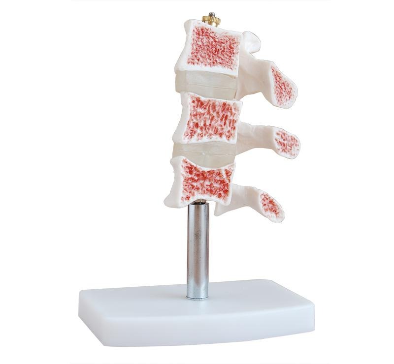
This anatomical model represents a cutaway view of the human vertebral column affected by osteoporosis. It clearly demonstrates the internal structure of the vertebrae and the gradual loss of bone density caused by osteoporosis. The porous red areas inside the vertebrae illustrate weakened trabecular bone, helping students and medical professionals understand the effects of bone degeneration. Mounted on a durable stand, this model is widely used in medical colleges, hospitals, nursing institutions, and biology laboratories for educational and demonstration purposes.
VVSSCIENTIFICS Products
919843643866
Chat with us 


This is your website preview.
Currently it only shows your basic business info. Start adding relevant business details such as description, images and products or services to gain your customers attention by using Boost 360 android app / iOS App / web portal.
Scientific And Laboratory Equipment In Salem
2026-05-26T06:55:39
This anatomical model represents a cutaway view of the human vertebral column affected by osteoporosis. It clearly demonstrates the internal structure of the vertebrae and the gradual loss of bone density caused by osteoporosis. The porous red areas inside the vertebrae illustrate weakened trabecular bone, helping students and medical professionals understand the effects of bone degeneration. Mounted on a durable stand, this model is widely used in medical colleges, hospitals, nursing institutions, and biology laboratories for educational and demonstration purposes.
2026-05-26T06:55:39


Submit Your Enquiry